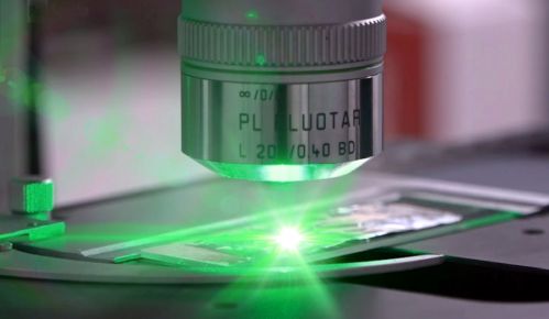
An international research team coordinated by IFN-CNR in Milan has developed a new optical microscope that can produce more efficiently than currently used systems detailed images of a sample’s chemical composition. This breakthrough opens up new possibilities in the fields of materials science and life sciences. The results, achieved in collaboration with the Politecnico di Milano, Columbia University, and Stanford University, have been published in the journal Optica.
Here the link to the original article: B. Ardini, A. Bassi, A. Candeo, A. Genco, C. Trovatello, X. Zhu, G. Valentini, G. Cerullo, R. Vanna and C. Manzoni, “High-throughput multimodal wide-field Fourier-transform Raman microscope”
“The instrument represents an important advancement in the field of microscopy and spectroscopy, opening up new perspectives for research in materials science and life sciences. It can contribute to the study of innovative two-dimensional materials and the detection and characterization of microplastics found in the environment and animal tissues,” says the research coordinator, Cristian Manzoni (CNR-IFN).
The benefits offered by the microscope stem from the novel combination of two techniques: Raman spectroscopy and Fourier transform spectroscopy.
The Raman effect is a well-established physical phenomenon used for decades to obtain information about the composition of a sample without disturbing it. It allows for the generation of two-dimensional maps of the properties of a material or biological tissue.
In the work published in the journal Optica, the researchers demonstrate, through Fourier transform spectroscopy, that they have reduced the time required to acquire a detailed image of the sample compared to the longer time taken with Raman microscopes. Raman microscopes measure a spectrum for each point by scanning its surface, which is a slow process taking approximately 1 second per point (pixel).
Fourier transform spectroscopy, on the other hand, enables the simultaneous measurement of all points on the sample, eliminating the spatial or spectral filters used in traditional techniques. This method, based on a technique called interferometry, combines high efficiency with the ability to acquire multiple data points on the same sample simultaneously.
In their study, the researchers employed an exceptionally stable and repeatable birefringent interferometer. The system acquires Raman and fluorescence maps with high spatial resolution (less than 1 micrometer) in a time up to 100 times faster than traditional instruments. “This method also allows for the separate measurement of Raman and fluorescence signals, enabling the unprecedented study of both phenomena in the same area of the sample and obtaining much more spectral information than traditional techniques,” concludes Manzoni.